What are the principles of MRI

Andrew Vasquez
Published May 21, 2026
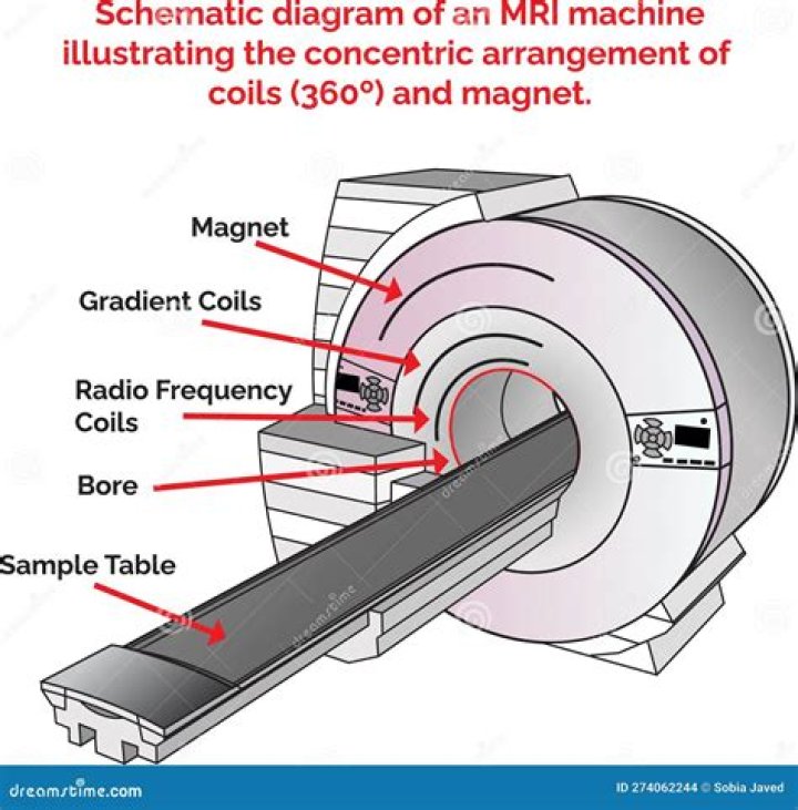
MRI machines are based on the same principle as are electromagnets, which produce a magnetic field by passing an electrical current through a massive coil. To eliminate electrical resistance, the coil is enveloped by liquid helium (−273 °C) to bring it into a superconducting state.
What is the basic principles of MRI?
Magnetic Resonance – Basic Principles The hydrogen nuclei behave like compass needles that are partially aligned by a strong magnetic field in the scanner. The nuclei can be rotated using radio waves, and they subsequently oscillate in the magnetic field while their magnetization returns to equilibrium.
What is the principle of MRI Class 10?
The principle used in MRI is that the protons in different types of human tissues have their own specific magnetic field environment.
What is the principle behind MRI give its application?
MRI scanners use strong magnetic fields, magnetic field gradients, and radio waves to generate images of the organs in the body. MRI does not involve X-rays or the use of ionizing radiation, which distinguishes it from CT and PET scans.What is the principle of CT scan?
CT uses ionizing radiation, or x-rays, coupled with an electronic detector array to record a pattern of densities and create an image of a “slice” or “cut” of tissue. The x-ray beam rotates around the object within the scanner such that multiple x-ray projections pass through the object (Fig 1).
What is matrix in CT scan?
Matrix: Two dimensional grid of pixels, used to compose images on a display monitor. The matrix determines the number of rows and columns. Partial volume effect: When different tissues/objects are represented by the same voxel. Each tissue/object only partially fills the voxel and is therefore a partial volume.
Which rays are used in MRI scan?
The MRI uses magnetic wave, whereas the X-ray uses radiation. They both can take pictures of the inside of the body and can be used for a better diagnosis of an injury or illness.
How is fMRI different from MRI?
What’s the Difference Between MRI and FMRI? FMRI scans use the same basic principles of atomic physics as MRI scans, but MRI scans image anatomical structure whereas FMRI image metabolic function. Thus, the images generated by MRI scans are like three dimensional pictures of anatomic structure.How much is a functional MRI?
How Much Does a Functional MRI (fMRI) Cost? On MDsave, the cost of a Functional MRI (fMRI) ranges from $488 to $623. Those on high deductible health plans or without insurance can save when they buy their procedure upfront through MDsave.
How does MRI measure brain activity?Functional magnetic resonance imaging or functional MRI (fMRI) measures brain activity by detecting changes associated with blood flow. This technique relies on the fact that cerebral blood flow and neuronal activation are coupled. When an area of the brain is in use, blood flow to that region also increases.
Article first time published onWhat is the difference between CT scan and MRI?
The biggest difference between MRI and CT scans is that MRIs use radio waves while CT scans use X-rays. Following are several others. MRIs are typically more expensive than CT scans. CT scans may be quieter and more comfortable.
What's a MRA test?
Magnetic resonance angiography–also called a magnetic resonance angiogram or MRA–is a type of MRI that looks specifically at the body’s blood vessels. Unlike a traditional angiogram, which requires inserting a catheter into the body, magnetic resonance angiography is a far less invasive and less painful test.
Who invented MRI?
Raymond Damadian, the inventor of the first magnetic resonance scanning machine celebrates his 85th birthday on March 16. Damadian, a physician, performed the first full-body scan of a human being in 1977.
What is better MRI or xray?
MRIs are more versatile, and doctors use them for examining many medical conditions. For example, x-rays are used more for examining broken bones, but they can also help detect diseased tissue. MRIs are better for evaluating soft tissues such as tendon and ligament injuries, brain tumors or spinal cord injuries.
Which is safer MRI or xray?
A. Magnetic resonance imaging, or M.R.I., is considered one of the safest technologies for looking deep inside the body, because it doesn’t carry the radiation risk of X-rays or PET scans.
Which is safer MRI or CT scan?
The biggest differences between an MRI and a CT Scan is the use of radiation and a magnetic field. An MRI does not use radiation, and a CT Scan does not use a magnet. Meaning, one is safer than the other for some patients.
Is MRI more detailed than CT?
A CT scan uses X-rays, whereas an MRI scan uses strong magnetic fields and radio waves. CT scans are more common and less expensive, but MRI scans produce more detailed images.
Why head CT scan is done?
Computed tomography (CT) of the head uses special x-ray equipment to help assess head injuries, severe headaches, dizziness, and other symptoms of aneurysm, bleeding, stroke, and brain tumors. It also helps your doctor to evaluate your face, sinuses, and skull or to plan radiation therapy for brain cancer.
Why do you need to drink water before a CT scan?
Preparing for a CT scan The water hydrates you prior to having contrast media for the CT. In the waiting area you will be asked to drink another 500ml of water which outlines the stomach and bowel clearly on the scans. The water also helps fill your bladder so that it shows on the scan.
What is the CT number of water?
a normalized value of the calculated x-ray absorption coefficient of a pixel (picture element) in a computed tomogram, expressed in Hounsfield units, where the CT number of air is -1000 and that of water is 0.
What is window width in CT?
The window width is the range of the grayscale that can be displayed. The center of grayscale range is referred to as the window level.
What is slice thickness in CT?
Slice thickness and slice increment are central concepts that surround CT/MRI imaging. Slice thickness refers to the (often axial) resolution of the scan (2 mm in the illustration). Slice Increment refers to the movement of the table/scanner for scanning the next slice (varying from 1 mm to 4 mm in the illustration).
Can MRI brain scans be wrong?
Yes, it is possible. In fact, a radiologist can misread an X-ray, mammogram, MRI, CT, or CAT scan. And it happens more often than you might think. This causes misdiagnosis or failure to diagnosis an existing issue.
Does insurance cover functional MRI?
Rad Alliance, Inc., and it’s affiliations have made this technology accessible to the public for clinical evaluations. However, the use of fMRI for most conditions is still considered by insurance companies to be research and investigational and therefore is not covered by insurance.
What is a PET scan brain?
A brain positron emission tomography (PET) scan is an imaging test of the brain. It uses a radioactive substance called a tracer to look for disease or injury in the brain. A PET scan shows how the brain and its tissues are working.
What is the difference between functional and structural imaging?
Structural imaging refers to approaches that are specialized for the visualization and analysis of anatomical properties of the brain. … In contrast, functional imaging is used to identify brain areas and underlying brain processes that are associated with performing a particular cognitive or behavioral task.
Which is better EEG or MRI?
In general, MRI is good at telling us where the lesion is, whereas EEG is good at separating normal and abnormal primarily cortical function. The topologic usefulness of EEG is limited, although it may be improved with computerization.
What scan shows brain activity?
A form of MRI known as functional MRI (fMRI) has emerged as the most prominent neuroimaging technology over the last two decades. fMRI tracks changes in blood flow and oxygen levels to indicate neural activity. When a particular brain area is more active, it consumes more oxygen, and blood flow increases.
What are the disadvantages of MRI?
Disadvantages of MRI: MRI cannot always distinguish between malignant tumors or benign disease (such as breast fibroadenomas), which could lead to a false positive results. MRI is not painful, but the patient must remain still in an enclosed machine, which may be a problem for claustrophobic patients.
What is better than an MRI?
MRI. CT scans are more widely used than MRIs and are typically less expensive. MRIs, however, are thought to be superior in regards to the detail of the image. The most notable difference is that CT scans use X-rays while MRIs do not.
Can MRI see neurons?
If MRI could measure brain activity at a columnar level, scientists might be able to use that to draw conclusions about computations in individual neurons. This would be exciting because one of the limitations of MRI is that it can’t measure neuronal activity directly.