Is plantar aponeurosis elastic

Jessica Wilkins
Published May 17, 2026
Elastic energy within the human plantar aponeurosis contributes to arch shortening during the push-off phase of running.
Is plantar fascia elastic?
The in vivo elastic properties of the plantar fascia during the contact phase of walking were determined experimentally by integrating a pressure-sensitive optical gait platform with a radiographic fluoroscopy system for recording skeletal motion.
Is plantar aponeurosis a tendon?
1997). In the long term, after fasciotomy, foot pain may also recur due to overpronation of the foot (Tweed Barnes & Allen, 2009). Plantar fasciitis is diagnosed on clinical grounds, possibly with the support of imaging to rule out any other diseases and confirm a thickening of the PF.
What is the plantar aponeurosis made of?
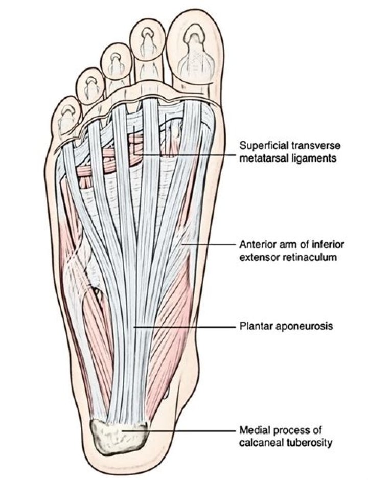
The Plantar aponeurosis is the modification of Deep fascia, which covers the sole. It is a thick connective tissue, that functions to support and protect the underlying vital structures of the foot. The fascia is thick centrally, known as aponeurosis and is thin along the sides.What is plantar fascia aponeurosis?
The plantar fascia is a thickened fibrous aponeurosis that originates from the medial tubercle of the calcaneus, runs forward to insert into the deep, short transverse ligaments of the metatarsal heads, dividing into 5 digital bands at the metatarsophalangeal joints and continuing forward to form the fibrous flexor …
Where do you get aponeurosis?
Aponeuroses are important for human movement and posture and are found all over your body, from the tip of your head to the soles of your feet. What, exactly, is an aponeurosis? An aponeurosis is a type of connective tissue that provides a point for a muscle to attach to a bone or cartilage.
What is an aponeurosis in anatomy?
aponeurosis, a flat sheet or ribbon of tendonlike material that anchors a muscle or connects it with the part that the muscle moves. The aponeurosis is composed of dense fibrous connective tissue containing fibroblasts (collagen-secreting spindle-shaped cells) and bundles of collagenous fibres in ordered arrays.
What are the attachments of the plantar fascia?
Arising predominantly from the calcaneal tuberosity, the plantar fascia attaches distally, through several slips, to the plantar aspect of the forefoot as well as the medial and lateral intermuscular septa. Anatomically, the fascia can be divided into three bands: the medial, lateral, and central.Where is the dorsum of foot?
The dorsum of foot is the area facing upwards while standing.
What supports the medial longitudinal arch?The plantar fascia is a major supporting element of the medial longitudinal arch that also plays a role in shock absorption.
Article first time published onIs the plantar fascia a tendon or ligament?
Plantar fasciitis (say “PLAN-ter fash-ee-EYE-tus”) is the most common cause of heel pain. The plantar fascia is the flat band of tissue (ligament) that connects your heel bone to your toes. It supports the arch of your foot. If you strain your plantar fascia, it gets weak, swollen, and irritated (inflamed).
What type of structure is the plantar fascia?
The plantar fascia is the thick connective tissue (aponeurosis) which supports the arch on the bottom (plantar side) of the foot. It runs from the tuberosity of the calcaneus (heel bone) forward to the heads of the metatarsal bones (the bone between each toe and the bones of the mid-foot).
Where is the plantar tendon?
The plantar fascia is a long, thin ligament that lies directly beneath the skin on the bottom of your foot. It connects the heel to the front of your foot, and supports the arch of your foot. The plantar fascia is a ligament that lies beneath the skin on the bottom of your foot.
What Innervates the plantar fascia?
The dorsal fascia is supplied by the superficial fibular nerve, the lateral fascia is supplied by the sural nerve, the medial fascia is supplied by the saphenous nerve, and the plantar fascia is supplied by the lateral and medial plantar nerves.
What is plantar aspect of foot?
The sole is the bottom of the foot. In humans the sole of the foot is anatomically referred to as the plantar aspect.
What is the plantar part of the foot?
The bottom of the foot is known as the sole. The padded area on the bottom of the foot is known as the plantar aspect.
What is the difference between palmar and plantar aponeurosis?
The palmar aponeuroses occur on the palms of the hands. … The plantar aponeuroses occur on the plantar aspect of the foot. They extend from the calcaneal tuberosity then diverge to connect to the bones, ligaments and the dermis of the skin around the distal part of the metatarsal bones.
What type of tissue is aponeurosis?
Aponeuroses are connective tissues found on the surface of pennate muscles and are in close association with muscle fascicles.
Why is aponeurosis different from tendon?
Key Difference The main difference is that Aponeurosis connects the muscles of the body to other muscles which necessitate help, while the tendons serve as a link between the muscles and the bones.
Are fascia and aponeurosis the same thing?
is that aponeurosis is (anatomy) a flattened fibrous membrane, similar to a tendon, that binds muscles together or connects them to other body parts like skin or bone while fascia is a wide band of material covering the ends of roof rafters, sometimes supporting a gutter in steep-slope roofing, but typically it is a …
Which is an example of an aponeurosis quizlet?
fascicles are inserted into the tendon from each side (from 2 sides).
Which of the following is an example or are examples of an aponeurosis?
It forms into sheets of pearly-white fibrous tissue to attach muscles needing a wide area of attachment. Aponeurosis can thin into a tendon and become a point of origin or insertion for other muscles. Some examples of aponeurotic fascia include the fascia of limbs, thoracolumbar fascia, and rectus sheath.
What Innervates the dorsum of the foot?
Dorsum skin is supplied by the terminal branches of tibial nerve and common peroneal nerve. Branches of the superficial peroneal nerve (SPN) supplies major portion of the dorsum of the foot and toes except the areas supplied by the deep peroneal nerve (DPN) and sural nerve (SN).
Which muscle is located in the dorsum?
Dorsal muscles of the foot. The muscles of the dorsum of the foot are a group of two muscles, which together represent the dorsal foot musculature. They are named extensor digitorum brevis and extensor hallucis brevis.
What is hand dorsum?
The dorsum of hand (opisthenar area, dorsal area) is the corresponding area on the posterior part of the hand.
Where do plantar fascia attach?
It involves inflammation of the plantar fascia — a tough, fibrous band of tissue that runs along the sole of the foot. The plantar fascia attaches to the heel bone (calcaneus) and to the base of the toes. It helps support the arch of the foot and has an important role in normal foot mechanics during walking.
Where is the medial band of the plantar fascia?
The medial and central bands originate from the larger medial calcaneal tubercle and fan forward toward the metatarsal heads and insert. The lateral band originates from the smaller lateral tubercle and fans distally to the plantar surface of the fifth metatarsal base.
How do you confirm plantar fasciitis?
Plantar fasciitis is diagnosed based on your medical history and physical examination. During the exam, your doctor will check for areas of tenderness in your foot. The location of your pain can help determine its cause.
What supports the transverse arch of the foot?
Transverse Arch The transverse arches are strengthened by the interosseous, plantar, and dorsal ligaments, by the short muscles of the first and fifth toes (especially the transverse head of the Adductor hallucis), and by the Peroneous longus, whose tendon stretches across between the piers of the arches.
What are plantar arches?
The plantar arch supplies the underside, or sole, of the foot. The plantar arch runs from the 5th metatarsal and extends medially to the 1st metatarsal (of the big toe).
What tendon supports the longitudinal arch of the foot?
The long plantar ligament bridges over, or rather under, the peroneus longus tendon – here’s the tendon, going to its insertion on the base of the first metatarsal. There’s another, even more impressive structure that supports the arch of the foot – the plantar aponeurosis.